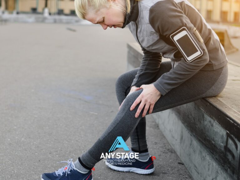
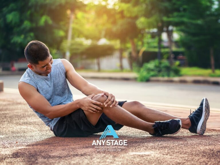
Jumper’s knee, also known as patellar tendinitis (indicates inflammation) or patella tendinopathy (may involve degenerative processes, failed healing responses, or a combination of both), is a common injury that affects the patellar tendon, which connects the patellar (knee cap) to the tibia (shin bone).
As the name suggests, Jumper’s Knee is a common complaint among athletes and people involved in plyometric sports such as, but not limited to, running, netball, basketball and volleyball.
Function and Structure of the Patellar Tendon (Jumper’s Knee)
 The patellar tendon starts on the patella (knee cap) and travels down the front of the knee to insert approximately 4.5cm (adults) below the knee cap on the tibia (tibial tuberosity). The patellar tendon shares fibres of the quadricep tendon, which attaches the rectus femoris (one of the quadricep muscles) to the patella. When the quadriceps muscles contract, the patellar tendon is pulled, which in turn straightens the knee and extends the leg. The patellar tendon’s role in transmitting forces during jumping indirectly influences the distribution of impact forces across the knee joint. As the quadriceps contract and the patellar tendon engages, it helps stabilise the knee joint and distribute forces more evenly, thereby reducing the stress on the surrounding soft tissues and minimising the risk of injury when jumping and landing.
The patellar tendon is surrounded by a tendon sheath, which helps to reduce friction between the tendon and the surrounding tissue. Blood vessels and nerves also run through the tendon sheath to supply the tendon with nutrients and sensory information.
Causes of Jumper’s Knee
Jumper’s knee is caused when tensile stress that is placed on the patellar tendon during activities that involve jumping or running, exceeds the capacity of the tendon. When the knee joint is extended, the quadriceps muscle pulls on the patellar tendon, which in turn places tension on the tendon fibres. This tension can cause microtrauma or small tears in the tendon, which can lead to pain, swelling, and inflammation.
Over time, if the tendon does not have time to heal properly or if the stress on the tendon continues, the microtrauma can progress to degeneration of the tendon tissue. This can result in a thickening of the tendon, decreased blood flow to the tendon, and increased pain and inflammation.
- Repetitive Stress: Continuous and repetitive jumping or landing activities can place excessive stress on the patellar tendon, leading to microtrauma and tissue damage over time. This repetitive stress is often a result of training or participating in sports that involve frequent jumping, cutting, or sudden changes in direction.
- Overuse: Jumper’s knee is typically considered an overuse injury, meaning it develops gradually due to repetitive stress on the tendon without adequate rest or recovery periods. Athletes who engage in high-intensity training without sufficient rest intervals may be at increased risk of developing patellar tendinopathy.
- Relative Weakness: Weakness, with respect to the demand on the knee, of the muscles surrounding the knee, particularly the quadriceps, hamstrings and gluteal complex, can alter biomechanics and increase the load placed on the patellar tendon during jumping and landing movements. Relative weakness or reduced flexibility may predispose individuals to develop a jumper’s knee.
- Biomechanics: Inefficient movement patterns, such as improper landing technique, excessive knee valgus (inward collapse), or overpronation of the feet, can contribute to increased stress on the patellar tendon during athletic activities. Poor biomechanics may result from inadequate coaching, improper training techniques, relative weakness or underlying structural abnormalities.
- Training Load: Rapid increases in training intensity, duration, or volume without appropriate progression or periodisation can overwhelm the body’s ability to adapt and lead to tissue breakdown. Training errors, such as sudden spikes in activity level, insufficient warm-up or cool-down, or inadequate recovery strategies, may predispose athletes to develop jumper’s knee.
- Footwear and Surface Factors: Inappropriate footwear, worn-out shoes, or training on hard surfaces with insufficient shock absorption can exacerbate stress on the patellar tendon during jumping and landing movements.
- Previous Injuries: Prior history of knee injuries, such as patellar tendinitis, ligament sprains, or meniscus tears, may predispose individuals to develop recurrent or chronic symptoms of jumper’s knee. Residual weaknesses, altered movement patterns, or unresolved tissue damage from previous injuries can contribute to ongoing patellar tendon dysfunction.
- Biological Factors: Certain anatomical or physiological factors, such as age, genetics, body composition, and hormonal influences, may influence an individual’s susceptibility to developing a jumper’s knee. Older athletes, adolescents experiencing growth spurts, and individuals with a family history of tendon disorders may be at increased risk.
Addressing these specific causes through comprehensive assessment, targeted rehabilitation strategies, and modification of training and biomechanics can help prevent and manage a jumper’s knee effectively. It’s essential for athletes to work with a Physiotherapist and coaching staff, to identify risk factors, implement appropriate interventions, and optimise performance while minimising the risk of injury

Common Symptoms of Jumper’s Knee
The most common symptoms of patellar tendonitis are:
- Pain during sports/active movements
- Sharp, throbbing pain beneath the knee cap, particularly at the start of the sport/activity event. It is common for the pain to reduce as the sport/activity continues. If left untreated, the pain may become more consistent and remain at rest.
- Pain just below the knee cap with activities such as jumping, running, squatting
- Pain with any pressure through the tendon e.g. touch, kneeling
- Aching and stiffness after activity
- Stiffness in the morning
- Thickening of the tendon
The symptoms of jumper’s knee tend to get worse over time, increasing in frequency and intensity.
What is Patellar Tendinopathy?
When the patellar tendon doesn’t have the appropriate amount of load, sufficient time to repair, or the stress on the tendon is too high for the tendon to manage, they can develop tendinopathy. Tendinopathy is a condition that refers to damage or degeneration of a tendon, which can cause pain, swelling, and reduced function. Tendinopathy can occur in various parts of the body, but it is most common in the Achilles tendon, rotator cuff tendons, and the patellar tendon.
There are typically three stages of tendinopathy:
Reactive stage:
- This initial stage of tendinopathy is characterised by tendon inflammation and a reactive response to excessive mechanical loading or repetitive stress.
- Symptoms may include localised pain, swelling, and stiffness, particularly during or after activity.
- Scans may reveal disorganisation of collagen fibres, increased vascularity, and infiltration of inflammatory cells.
- At this stage, the tendon retains its structural integrity, and there is potential for recovery with appropriate management, including relative rest, activity modification, and rehabilitation.
Disrepair stage:
- There is a failed attempt at tissue healing characterised by disorganised collagen synthesis and remodelling.
- Microscopic changes in the tendon tissue may include increased cellularity, collagen disorganisation, and the presence of abnormal matrix components.
- The tendon may exhibit structural alterations, such as focal areas of thickening or nodularity, reflecting ongoing tissue remodelling processes.
- Management strategies at this stage may focus on promoting appropriate tendon healing through targeted exercise interventions, load management, and biomechanical optimisation.
- This stage can last for several weeks to several months.
Degenerative stage:
- The degenerative tendinopathy stage represents advanced tendon pathology characterised by irreversible structural changes and tissue degeneration.
- Symptoms may become chronic and debilitating, with persistent pain, functional limitations, and reduced tolerance to activity.
- Microscopic examination of the tendon reveals extensive collagen disorganisation, fibre degeneration, focal necrosis, and the accumulation of abnormal matrix substances.
- Imaging studies, such as ultrasound or MRI, may show evidence of tendon thickening, intratendinous tears, and calcifications.
- Treatment options for degenerative tendinopathy are aimed at symptom management, improving function, and preventing further deterioration. These may include modalities such as eccentric exercise therapy, shockwave therapy, corticosteroid injections (in select cases), and surgical intervention.
It is important to note that not all cases of tendinopathy progress through all three stages, and the severity and duration of symptoms can vary depending on the individual and the type of tendon involved.

Treatment options for Jumper’s Knee
Effective treatment for jumper’s knee typically involves a combination of relative rest from the aggravating activity, specific load management, strengthening exercises, physiotherapy-guided rehabilitation, and other interventions such as ice, compression, and anti-inflammatory medication.
Rehabilitation exercises focused on strengthening the quadriceps, hamstring, gluteal complex and calf muscles can help to improve the biomechanics of the knee and reduce stress on the patellar tendon. In some cases, more aggressive treatments such as corticosteroid/platelet-rich plasma (PRP) injections or surgery may be necessary.
Exercise type:
The type of exercise to perform is determined by the stage of injury and irritability of the tendon and progresses in the following order:
Isometric exercises
Isometric exercises of the quadricep involve contracting the muscle without moving the joint. These exercises can help strengthen the quadriceps muscles without putting additional stress on the patellar tendon.
Examples include:
- Quad sets: Sit with your legs straight out in front of you and your back against a wall. Tighten your thigh muscles and press the back of your knee into the floor for 5-10 seconds, then release.
- Wall sits: Lean your back against a wall and slide down until your knees are bent at a 90-degree angle. Hold this position for 30-45 seconds, then stand back up. Depth and endurance will be determined by irritability of the patellar tendon.
Eccentric exercises
Eccentric exercises involve lengthening the muscle while it is contracting, which can help improve tendon strength and reduce pain.
Examples include:
- Single-leg decline squats: Stand on a step or platform with your heels hanging off the edge. Slowly lower your body down until your knees are bent at a 90-degree angle, then push back up using both legs. Repeat for several reps on each leg.
- Eccentric step-ups: Stand with one foot on a step or platform and the other foot on the ground. Slowly lower your body down until your front knee is bent at a 90-degree angle, then push back up using both legs. Repeat for several reps on each leg.
Isotonic exercises
Isotonic exercises incorporate a concentric phase (muscle shortening) and eccentric phase (muscle lengthening), lifting a constant load throughout the range of motion. These exercises are commonly used to improve muscle strength, endurance, and hypertrophy (muscle growth).
Examples include:
- Repeated knee extension: Sit on a chair with your feet on the floor. Straighten your leg out in front of you and pause, before slowly lowering your leg to the starting position. Repeat 3×12. Progressions include adding weight and performing it with a single leg.
- Squats: Start by standing with your feet slightly outside shoulder width. Place a chair behind you for guidance. Slowly bend your knees and take your hips back toward the chair, as if you were going to sit down. Lightly touch the chair, before standing up and returning to the starting position. Repeat 3×12. Progressions include removing the chair sitting deeper and adding weight.
Stretching exercises
Tight muscles around the knee can contribute to jumper’s knee. Stretching exercises can help improve flexibility and reduce tension.
Examples include:
- Hamstring stretch: Sit with your legs straight out in front of you and your back against a wall. Reach forward and try to touch your toes, holding the stretch for 15-30 seconds.
- Standing Quad Stretch: Stand upright with feet hip-width apart. Shift your weight onto one leg and maintain your balance. Bend your other knee, bringing your heel toward your buttocks. Reach back with the hand on the same side as the bent knee and grasp the ankle or foot. Gently pull your heel closer to your buttocks, feeling a stretch along the front of the thigh. Keep your knees close together and your torso upright, avoiding leaning forward or arching the lower back excessively. Hold the stretch for 20-30 seconds, breathing deeply and maintaining a comfortable level of tension. Release the stretch slowly and repeat on the opposite side. This can cause compression of the patellar tendon. Be sure to hold the stretch without irritating the tendon.
- Calf stretch: Stand facing a wall with one foot in front of the other. Lean forward and place your hands on the wall, keeping both heels on the ground. Hold the stretch for 15-30 seconds, then switch legs.

Increasing Tendon Capacity
Increasing the capacity of the patellar tendon involves the strengthening of surrounding muscle groups and the ability to tolerate sports/activity demands. This includes proper training techniques, such as gradually increasing the intensity and frequency of activity and using proper landing and jumping techniques. Maintaining adequate flexibility and strength in the quadriceps, hamstrings, and calf muscles can also help to prevent the development of jumper’s knee.
The information provided above is general in nature. If you have experienced jumper’s knee, or, you have persistent knee pain, please do not hesitate to contact our friendly and experienced team at Any Stage Physiotherapy and Sports Medicine for individualised assessment and information.
We are here to guide you back to a pain-free lifestyle and help return you to sport.